95.2.-HYDATID CYSTS IN SOUTHWEST OF SPAIN. A FORENSIC POINT OF VIEW. HISTOPATHOLOGICAL FINDINGS IN LUNGS AND LIVER.
Prof. Garfia.A
95.2-HIDATIDOSIS EN PATOLOGÍA FORENSE EN EL SUROESTE DE ESPAÑA: HALLAZGOS PULMONARES Y HEPÁTICOS.
Prof.Garfia.A
95.2-hydatidosis IN FORENSIC PATHOLOGY IN SOUTHWEST SPAIN: PATHOLOGICAL FINDINGS IN LIVER AND LUNG.
Prof.Garfia.A
CASO N º 2 -
NOTA
Las lesiones granulomatosas, detected in the lung in this case, be regarded as an evolutionary morphological stage in the development of scolices planted in the lung, following the rupture of a hydatid cyst, as was the case previously described (see case: 94.1).
Case description
Male, 58 years old, resident of a people of Extremadura, retired hunter, who was engaged in farming and working with dogs, rabbits and foxes.
Circumstances of death
The man underwent surgery with general anesthesia, to present a loose body in the right elbow joint. During the operation cardiac arrest, which was followed by the death, despite the application of advanced resuscitation.
Family challenge conducted a judicial autopsy, considering death occurred during surgery minor surgery.
Procolo of elective surgery .-
After the radiological diagnosis of articular loose body right elbow, is scheduled surgery. Were conducted the following analysis complementary, before the intervention:
AP 1.-Plain radiograph of the chest. was considered within normal limits.
2.-Hematology, presented the following:
Leukocyte ........ 6,600 ml.
......... 4.73 million red cells ml.
15.3 ................... Hb g.
Hcm ................ 44.2%.
Vcm ................. 92.
........ Platelets 190,000
bleeding time ... 1m. 45 "
prothrombin T. ....... 92%.
TTP ........... .. 29 "
WBC count .... not practiced.
Glycemic ...... 93 mg / dl.
............. Urea 31 mg / dl.
............... Normal ECG.
PA 14 / 9
Frequency cardiaca...64/min, the day of admission.
PART OF ANESTHESIA
Preoperative .- C aken within the normal range for their age.
Intervention .-
peripheral cannulation Abbocath track of the number 18, ECG monitoring is required; atropinization and oral intubation, after ventilation tube, mechanical ventilation is changed . Normal lung sounds and MT 14 / 9.
A few minutes into the intervention, immediately after incision of the skin and subcutaneous tissue is bradycardia occurred, with complete atrioventricular dissociation, and the establishment of peripheral cyanosis. We proceeded to the administration of oxygen, 100%, and atropine, without obtaining a reply. Given Dacorsol and Aleudrina, lowering the heart rate to 30 s / min, followed by stop Aleudrina attempted to trace and external cardiac massage, heart rate recovered to 50 s / min and began to appear premature ventricular contractions, passing to tachycardia and ventricular fibrillation. Cardioversion is applied; appearance of idioventricular rhythm, and new atrial fibrillation. Temporary pacemaker was installed with the patient in mydriasis and cyanosis. The patient was exitus.
was performed a forensic autopsy, due to the claim of malpractice family.
Autopsy Findings
External Review
is the body of a male, athletic build, Cadaveric phenomena presented in accordance with the time of death. Cervico-facial cyanosis and a bandage on his right elbow in the surgical incision site.
Internal Review
existed pleural adhesions, bilateral, with little pathological significance, as well as the presence of subpleural ecchymoses. The lung was congestive and edematous. The coroner who performed the autopsy did not detect the presence of any cyst ruptured, as in the previous case (94.1). heart, 550 g, had free patent coronary atherosclerotic plaque. In the epicardium were detected focal hemorrhages attributed to CPR. The blood was cherry red.
Cause obituary in the autopsy examination:
Death by cardiac arrest during surgery, accidental cause.
HISTOPATHOLOGY
were issued the following histopathological diagnoses:
Heart .-
cardiac hypertrophy (increase weight percentage of 50%, aproximadadamente considering a weight limit of 340 g, for a adult in our country).
concentric left ventricular hypertrophy . Hypertensive cardiomyopathy. Of coronary intramural arteriolosclerosis.
Liver .-
intense yellow color. Macro and microvesicular steatosis, de distribución periportal y mediozonal, de grado moderado.
Pulmón.-
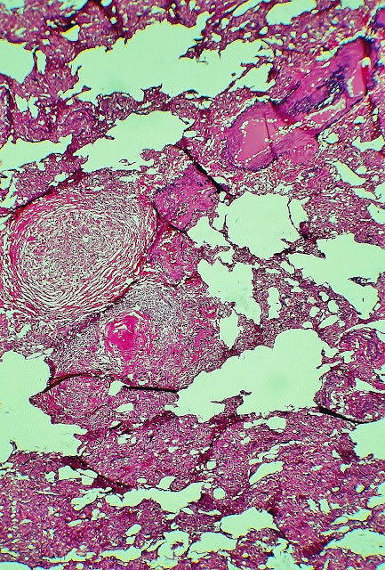
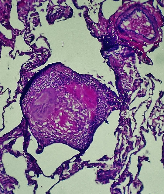
Numerosos granulomas, de tamaño variable entre 300-400 micras y varios mm, algunos de los cuales presentan reacción gigantocelular y respuesta linfocitaria.Periféricamente aparecen rodeados por una envoltura formada por gruesas fibras de colágeno, de distribución concéntrica. Los granulomas se encontraban en diferentes estadíos de desarrollo hacia la fibrosis.
DIAGNOSTIC Morphological characteristics of the hydatid cyst in forensic pathology.
hidatítico lung cyst is a parasitic cyst formation that forms in the lung where it nests in the embryonic form of the tapeworm echinococcus, whose adult form parasite, especially the dog. Is the pulmonary form of the disease equinococócica general.
Synonymy .- lung hydatid cyst, pulmonary echinococcosis, hydatidosis, hydatid echinococcosis. History .-
The true origin of hydatid disease is due to the work of Hartmann and Pallas. The pulmonary, and study, as Laennec was made out of several French writers. The modern concept of disease is due to Devi, who by the year 1917 made a comprehensive study has been done clásico.Por the high frequency of this disease seen in Argentina, American authors have studied carefully (Escudero published in excellent monograph 1912, coinciding with the work roentgeniano study initiation hydatid cyst, which was so fruitful for American authors such as Piaggio Blanco and Garcia Capurro, who, in 1939, published an excellent monograph on the subject.)
Etiology and Pathogenesis .-
hydatid cyst corresponds to one phase of the life cycle of Echinococcus granulosus. This parasite shows in their life cycle, two distinct phases: one larva, with formation of cysts in the intermediate host-usually sheep, lambs, and other herbivores, and a phase of adult worm, which occurs in dogs and other canine species. In this cycle of the parasite, man acts as an intermediate host, but without the possibility of transmission to the definitive host. The cycle begins when an animal, usually the group of dogs, ingested cysts present in the guts from a dead herbivore. Upon reaching the intestine, scolices attach to the wall of that, developing its adult form, which consists of a head or scolex and four rings / segments or proglottids. The total length of the adult tapeworm reaches between 4 and 8 mm. Eggs in the last proglottid are released into the intestinal lumen and removed later by faeces, thus producing water pollution and vegetables irrigated with it. The intermediate host, including humans, eat the eggs that hatch in the small intestine. The embryos through the intestinal wall and reach the venous drainage, reaching the liver, primarily, and also the lungs and other organs. The parasite settles in one of the above bodies and the host responds with a fibrous reaction in the periphery. Of a progressive, albeit slow, start the development of hydatid cyst that after several months, it can reach 10-20 cm in diameter.
Macroscopic appearance .-
Microscopic Morphology .-
The result of this evolutionary process is the formation of a cyst, surrounded by a layer of fibrous tissue generated by the host. This layer, called adventitious or pericyst layer, is around the cyst but not interact with him. The cyst wall, itself, is composed of two layers: 1) the innermost layer is the germ layer, or endoquiste prolígera membrane, is formed by a syncytium of a single cell layer and 2) a thick white membrane - formed by parallel plates of hyaline material, which are enucleated called laminated membrane, cuticular layer, or ectoquiste, originating from the germinal layer.
From the buds grow germ layer that transforms into stalked daughter cysts, the walls of the daughter vesicles invaginate to form scolices or protoscoleces.
parasite cycle ends when the intermediate host dies and some viscera parasitized is ingested by a carnivore, in general, the group of dogs.
the morphological diagnosis of a hydatid cyst SURE, in tissue, is based on THE PRESENCE OF ANY OF THE FOLLOWING COMPONENTS:
scolices 1.-infectious.
2.-hooks in the crown of a scolex.
3.-laminated membrane fragments.
NOTE
The series of photographs shown below were made with techniques silver impregnation (silver methenamine), hematoxylin-eosin-phloxine, staining for elastic fibers and PAS. The photo sequence shown attempts to indicate the different cuts examined to reach the accurate diagnosis of pulmonary hydatid disease. The morphological data target for the diagnosis was the presence of laminated hydatid membrane cuticle layer, or remnants of it, in some of the granulomas shown in recent photographs.
F ig. 1.-Lung. This photo and the following are from serial sections stained with different techniques histology. Sobretinción PAS technique and with elastic. The picture shows the amorphous, PAS positive located in the center of both granulomas. The top granuloma located in the vicinity of a bronchiole. Prof.Garfia.A
Fig.1.A-Lung. In the center of both granulomas distinguishes amorphous PAS positive material. Prof.Garfia.A
Procolo of elective surgery .-
After the radiological diagnosis of articular loose body right elbow, is scheduled surgery. Were conducted the following analysis complementary, before the intervention:
AP 1.-Plain radiograph of the chest. was considered within normal limits.
2.-Hematology, presented the following:
Leukocyte ........ 6,600 ml.
......... 4.73 million red cells ml.
15.3 ................... Hb g.
Hcm ................ 44.2%.
Vcm ................. 92.
........ Platelets 190,000
bleeding time ... 1m. 45 "
prothrombin T. ....... 92%.
TTP ........... .. 29 "
WBC count .... not practiced.
Glycemic ...... 93 mg / dl.
............. Urea 31 mg / dl.
............... Normal ECG.
PA 14 / 9
Frequency cardiaca...64/min, the day of admission.
PART OF ANESTHESIA
Preoperative .- C aken within the normal range for their age.
Intervention .-
peripheral cannulation Abbocath track of the number 18, ECG monitoring is required; atropinization and oral intubation, after ventilation tube, mechanical ventilation is changed . Normal lung sounds and MT 14 / 9.
A few minutes into the intervention, immediately after incision of the skin and subcutaneous tissue is bradycardia occurred, with complete atrioventricular dissociation, and the establishment of peripheral cyanosis. We proceeded to the administration of oxygen, 100%, and atropine, without obtaining a reply. Given Dacorsol and Aleudrina, lowering the heart rate to 30 s / min, followed by stop Aleudrina attempted to trace and external cardiac massage, heart rate recovered to 50 s / min and began to appear premature ventricular contractions, passing to tachycardia and ventricular fibrillation. Cardioversion is applied; appearance of idioventricular rhythm, and new atrial fibrillation. Temporary pacemaker was installed with the patient in mydriasis and cyanosis. The patient was exitus.
was performed a forensic autopsy, due to the claim of malpractice family.
Autopsy Findings
External Review
is the body of a male, athletic build, Cadaveric phenomena presented in accordance with the time of death. Cervico-facial cyanosis and a bandage on his right elbow in the surgical incision site.
Internal Review
existed pleural adhesions, bilateral, with little pathological significance, as well as the presence of subpleural ecchymoses. The lung was congestive and edematous. The coroner who performed the autopsy did not detect the presence of any cyst ruptured, as in the previous case (94.1). heart, 550 g, had free patent coronary atherosclerotic plaque. In the epicardium were detected focal hemorrhages attributed to CPR. The blood was cherry red.
Cause obituary in the autopsy examination:
Death by cardiac arrest during surgery, accidental cause.
HISTOPATHOLOGY
were issued the following histopathological diagnoses:
Heart .-
cardiac hypertrophy (increase weight percentage of 50%, aproximadadamente considering a weight limit of 340 g, for a adult in our country).
concentric left ventricular hypertrophy . Hypertensive cardiomyopathy. Of coronary intramural arteriolosclerosis.
Liver .-
intense yellow color. Macro and microvesicular steatosis, de distribución periportal y mediozonal, de grado moderado.
Pulmón.-
Numerosos granulomas, de tamaño variable entre 300-400 micras y varios mm, algunos de los cuales presentan reacción gigantocelular y respuesta linfocitaria.Periféricamente aparecen rodeados por una envoltura formada por gruesas fibras de colágeno, de distribución concéntrica. Los granulomas se encontraban en diferentes estadíos de desarrollo hacia la fibrosis.
DIAGNOSTIC Morphological characteristics of the hydatid cyst in forensic pathology.
hidatítico lung cyst is a parasitic cyst formation that forms in the lung where it nests in the embryonic form of the tapeworm echinococcus, whose adult form parasite, especially the dog. Is the pulmonary form of the disease equinococócica general.
Synonymy .- lung hydatid cyst, pulmonary echinococcosis, hydatidosis, hydatid echinococcosis. History .-
The true origin of hydatid disease is due to the work of Hartmann and Pallas. The pulmonary, and study, as Laennec was made out of several French writers. The modern concept of disease is due to Devi, who by the year 1917 made a comprehensive study has been done clásico.Por the high frequency of this disease seen in Argentina, American authors have studied carefully (Escudero published in excellent monograph 1912, coinciding with the work roentgeniano study initiation hydatid cyst, which was so fruitful for American authors such as Piaggio Blanco and Garcia Capurro, who, in 1939, published an excellent monograph on the subject.)
Etiology and Pathogenesis .-
hydatid cyst corresponds to one phase of the life cycle of Echinococcus granulosus. This parasite shows in their life cycle, two distinct phases: one larva, with formation of cysts in the intermediate host-usually sheep, lambs, and other herbivores, and a phase of adult worm, which occurs in dogs and other canine species. In this cycle of the parasite, man acts as an intermediate host, but without the possibility of transmission to the definitive host. The cycle begins when an animal, usually the group of dogs, ingested cysts present in the guts from a dead herbivore. Upon reaching the intestine, scolices attach to the wall of that, developing its adult form, which consists of a head or scolex and four rings / segments or proglottids. The total length of the adult tapeworm reaches between 4 and 8 mm. Eggs in the last proglottid are released into the intestinal lumen and removed later by faeces, thus producing water pollution and vegetables irrigated with it. The intermediate host, including humans, eat the eggs that hatch in the small intestine. The embryos through the intestinal wall and reach the venous drainage, reaching the liver, primarily, and also the lungs and other organs. The parasite settles in one of the above bodies and the host responds with a fibrous reaction in the periphery. Of a progressive, albeit slow, start the development of hydatid cyst that after several months, it can reach 10-20 cm in diameter.
Macroscopic appearance .-
The gross appearance of the hydatid cysts depends on the infecting species and the patient's immune response.
dependent cysts of Echinococcus granulosus larvae can reach a diameter of 10-20 cm and are encapsulated lesions, and well defined, containing a liquid, clear and transparent, water-like rock called hydatid fluid. Cysts can be located anywhere in the lungs. When the cyst reaches a diameter of about 20 cm, originating from the inside of your wall outgrowths (daughter cysts) containing scolices. The daughter cysts can break off and fall to the bottom of the cyst forming the so-called hydatid sand. "
Microscopic Morphology .-
The result of this evolutionary process is the formation of a cyst, surrounded by a layer of fibrous tissue generated by the host. This layer, called adventitious or pericyst layer, is around the cyst but not interact with him. The cyst wall, itself, is composed of two layers: 1) the innermost layer is the germ layer, or endoquiste prolígera membrane, is formed by a syncytium of a single cell layer and 2) a thick white membrane - formed by parallel plates of hyaline material, which are enucleated called laminated membrane, cuticular layer, or ectoquiste, originating from the germinal layer.
From the buds grow germ layer that transforms into stalked daughter cysts, the walls of the daughter vesicles invaginate to form scolices or protoscoleces.
parasite cycle ends when the intermediate host dies and some viscera parasitized is ingested by a carnivore, in general, the group of dogs.
the morphological diagnosis of a hydatid cyst SURE, in tissue, is based on THE PRESENCE OF ANY OF THE FOLLOWING COMPONENTS:
scolices 1.-infectious.
2.-hooks in the crown of a scolex.
3.-laminated membrane fragments.
NOTE
The series of photographs shown below were made with techniques silver impregnation (silver methenamine), hematoxylin-eosin-phloxine, staining for elastic fibers and PAS. The photo sequence shown attempts to indicate the different cuts examined to reach the accurate diagnosis of pulmonary hydatid disease. The morphological data target for the diagnosis was the presence of laminated hydatid membrane cuticle layer, or remnants of it, in some of the granulomas shown in recent photographs.
F ig. 1.-Lung. This photo and the following are from serial sections stained with different techniques histology. Sobretinción PAS technique and with elastic. The picture shows the amorphous, PAS positive located in the center of both granulomas. The top granuloma located in the vicinity of a bronchiole. Prof.Garfia.A
Fig.1.A-Lung. In the center of both granulomas distinguishes amorphous PAS positive material. Prof.Garfia.A
Fig.3.-eosin Pulmón.Hematoxilina floxina.Mostrando granuloma in the vicinity of a glass. Prof.Garfia.A
Fig.4.-Detail of the fibrous capsule granulomatous, evidenced with silver-methenamine. Prof.Garfia.A
Fig.5. "It's a small granuloma located next to an irregular space of the airway. Silver metenamina.Prof.Garfia.A
Fig.6.-Detail of Fig anterior.Corte stained with hematoxylin-eosin, elastic deVerhoeff. Prof.Garfia.A
Fig.7.-Two granulomas, one of which contains hyaline material in the center. Hematoxylin-eosin-phloxine. Prof.Garfia.A
Fig.8 .- The first granuloma in which it was observed the presence of multinucleated giant cells and a lymphocytic corona. Hematoxylin-eosin-elastic.
Fig 9.-Granuloma "soft", consisting of eosinophilic material remains, the presence of multinucleated giant cells and epithelioid cells. Prof.Garfia
Fig.10 .- Granuloma adjacent to the wall of a bronchiole, material containing amorphous eosinophilic center. Its location is suggestive of hydatid cyst bronchogenic dissemination. In certain cases has raised the differential diagnosis of granulomatosis bronchocentric (bronchocentric granulomatosis). Prof.Garfia.A
Fig.11.-up to the same cut-metenamina.Prof.Garfia.A silver stained
Fig.12.-Granuloma in the center containing eosinophilic material, suggesting amorphous, with the photograph, the presence of a parásito.Prof.Garfia.A
Fig.13 .- methenamine silver, the same court distinguished the present material, red brick, shaped brackets, very suggestive hydatid cyst, surrounding a mass of amorphous material in the center of granuloma.Prof.Garfia.A
Fig.14.-This photograph and the next show granulomas containing material strange, at its center, which adopts a morphology in "sailboat." There is no morphologic diagnostic criteria of certainty, hidatídico.Prof.Garfia.A cyst
Fig.15.-Detail of Fig anterior.Prof.Garfia.A
Fig.16 .- Granuloma containing membrane remnants of germinative membrane laminated inactiva.Prof.Garfia.A
Fig. 17 .- This was the first granuloma which could note that had remnants of laminated membrane and membrane germination inactiva.Prof.Garfia.A
Fig.18 .- Granuloma stained with Masson trichrome, presented remnants of the laminated membrane and central necrotic areas containing necrotic vesicles. Prof.Garfia.A
Fig.19.-Detail anterior.Prof.Garfia.A
Fig.20.-Review polarized light of a granuloma containing four quistes.Prof.Garfia.A
Fig.21 .- Pulmón.Plata-methenamine. The photograph shows four cysts encompassed by the same cover of dense collagen fibers orientado.En the center of each granuloma lamellar fragments are distinguished. Prof.Garfia.A
Fig.22.-Pulmón.Plata-methenamine. Granuloma containing small inactive hydatid cyst. Prof.Garfia.A
Fig.23 .- Detail of the previous figure showing the cover laminated and germinative membrane or prolígera, detached from aquella.Plata-methenamine. Prof.Garfia.A
Fig.24 .- Large increase showing the morphology of the granuloma in which you can see, clearly, the laminated structure of the cuticle or outer layer of the cyst wall and detachment of the germinative layer of the cyst into the light "inactive." Silver-methenamine . Prof.Garfia.A
Fig 25.-The photograph was made on a histological without dewaxing, in order to visualize, with Nomarski interference microscopy, the structure "as a highway" of the laminate hydatid cyst. The presence of the laminated membrane is a unique morphological feature of hydatid cysts. Prof.Garfia.A


0 comments:
Post a Comment