Wednesday, March 30, 2011

Does Walmart Vision Accept Avesis

Shorts + blazer again!


look Total:
Zara Boots and belt: Vintage.


(The sorry if there are not many photos and are not the best quality, but it was a "minirrepor" (see palabrejaa) to any noise ! Sorry! = ().


cieliness
Hello!

this spring Definitely for my clothes are shorts fetish high waist combined with a blazer either smooth or checkered eg, as in this case. In fact, you can also check the previous post, in which not separate myself from these two items ... to see if I passed and the fever ... but I doubt it, because I think a garment shorts super versatile either day or night, you can combine with any outfit and also stylized and greatly promote our set. And as the Blazer, as it is an elegant piece that can be combined in any way and always give a touch of chic to any look!

And you what, you are also a die-hard fans of the shorts and blazers? =)

I really am very sorry to have so abandoned the blog, but I'm super lack of time and most days between faculty, sports, nap (lol), practices .... I have no time to make me a picture or even the look is appropriate for the upload! jajajjaja I'm really sorry, I'll try again auqnue pace before and update more often!
By the way, I am very sorry that people who do not like how it will decide to stop her blog to be followers, but also appreciate it because I like people to be critical and be with me because she likes the blog, I do not make me a follower of your blog too! Han 2 low been giving me grief, but to see if I get more batteries!

And of course, I have to thank all the friendly bloggers who comment me more or less always in the post, much love I have for you because you are a soless and know that thanks to you is my dream to make every blog day gets a little older and go to best! Graciassss Million!

And nothing I say goodbye, I appear much, but when I do not stop splitting! hahahaha that you spend good semanaaa!
MUAAAA


Marya *

South Park Slash Doujinshi

97.4.-ECHINOCOCCOSIS IN FORENSIC PATHOLOGY IN SPAIN. PROF.GARFIA.A

97.4.-HYDATID CYSTS  IN SOUTHWEST OF SPAIN.  A FORENSIC POINT OF VIEW.  HISTOPATHOLOGICAL FINDINGS IN LUNGS AND LIVER.
Prof.Garfia.A

97.4-HIDATIDOSIS Patologia FORENSICS EN EN EL SURO DE ESPAÑA: HALLAZGOS Pulmonary Y HEPÁTICOS.
Prof.Garfia.A

97.4-hydatidosis IN FORENSIC PATHOLOGY IN SOUTHWEST SPAIN : PATHOLOGICAL FINDINGS IN LIVER AND LUNG.
Prof.Garfia.A


CASE No. 4
personal history
Patient 65, who has a history of allergy to the "straw." Smoker until a few years ago. Malta has experienced fever. Suffering from COPD and hypertension blood laying in drug treatment. He was referred to the Service of Digestive Surgery, from Internal Medicine, where he was examined and diagnosed with liver hydatid cyst of 20 cm plant diámetro.Fue admitted to undergo elective surgery.
Physical
Obesity. Normal color of skin and mucous membranes.
AP: decreased breath sounds in the right lung base. AC: No changes.
Investigations
Analysis: Leukocytes: 7,500 (Seg.64; Lin. 20; Mon.7; Eo.6; Bas.0.6). Hb: 15.8, Ht: 48.6; VCM : 90.2; HCCM: 32.5; PLQ. 226,000.
INR: 1.01, prothrombin time: 99%; T. cephalin: 40.2, fibrinogen: 393.
Glu: 107; Urea: 56; uric acid: 8.3, Albumin 4.4, Total Protein: 8.2, Creatinine: 1.22, LDH: 291; Amylase: 45, GOT: 21; GPT: 20; F.alcalina : 310; Bil.total: 0.58; Bil. Direct: 0.19; Cholinesterase: 11941, Cholesterol 231, Triglycerides: 113, Ca: 9.78, Na: 141, K: 4.5.
HBs Ag and HCV Ab: negative.
hydatidosis Serology: positive (1 / 400).
Rx Chest right hemidiaphragm elevation.
Rx abdomen Hepatomegaly .


.- Surgical Intervention
was conducted bilateral subcostal laparotomy, extended to last right intercostal space. Hydatid cyst is discovered that measures 25x20 cm. Occupies the entire right lobe and presents intimate and firm adhesion to the diaphragm, retroperitoneum, inferior vena cava, from the upper renal pole to the diaphragmatic hiatus, moved to the gallbladder and covers the posterior capsule-in-a right hepatic veins, which are linked to empties into the inferior vena cava. It makes quistoperiquistectomía closed after ligation of vascular structures and bile, cholecystectomy and resection of tongue remnant of the right lobe liver. It left two silicone drains subdiafragmático.Hemostasis satisfactoria.Cierre liver cell layers.


Fig. 1. - abdominal ultrasound.
was reported by the radiologists as compatible with hepatic hydatid cyst 20 cm in diameter.


FIG.2. -
abdominal CT. Report.
right hepatic lobe atrophy secondary to the presence of a cystic lesion measuring 21x16x20 cm. The lesion has a uniform capsule, with some punctate calcification therein. Inside the lesion is small objectify linear images that could correspond to membranes. After contrast administration, no evidence of uptake within it. The lesion is suggestive of hydatid cyst and is in contact with hepatic veins and intrahepatic segment of the inferior vena cava. In the remaining parenchymal focal no lesions. Normal-sized bile ducts. Spleen, pancreas and splenoportal axis, no pathological findings. Normal kidneys.


Fig. 3 .-
.- Surgical Intervention
It held a bilateral subcostal laparotomy, extended to last right intercostal space. Hydatid cyst is discovered that measures 25x20 cm.


Fig .4 .- cyst or cupa entire right lobe and has adhesions, intimate and firm, diaphragm, retroperitoneum, inferior vena cava, from the upper renal pole to the diaphragmatic hiatus, moved to the gallbladder biliary and the posterior includes, in its capsule-a right hepatic veins, which are linked to the opening in the inferior vena cava.


Fig.5 .- closed quistoperiquistectomía is performed after ligation of vascular structures and bile Tongue cholecystectomy and resection of the right lobe liver remaining. It left two silicone drains and subdiaphragmatic liver cell. Hemostasis satisfactoria.Cierre layers.


POST .-

Postoperatively, the patient has hypotension, severe bradycardia, and subsequent cardiac arrest. It began cardiopulmonary resuscitation associated with cardioactive drugs. The patient left the unemployment supraventricular tachycardia that was treated medically with pressors and oxygen saturation. Presents new bradycardia and subsequent cardiac arrest. Restarted resuscitation without success.
was performed an autopsy familiar legal malpractice complaint.


HISTOPATHOLOGY
1 .- .- Macroscopic description
; Macroscopically it was a giant cyst formation, spherical, 5 kg of weight, measuring 24 cm in diameter after fixation in formalin. The outer cyst surface had yellowish brown color, cut, cautioned a single cavity completely occupied by gelatinous material, yellowish-white, within which identified a laminar formation, blackish brown, friable and necrotic, 0.3 cm thick and 15 cm in length. The cyst wall had firm, whitish and about 1 cm thick, on the inside of the capsule could distinguish some areas of thickening and sinuous surface, the largest of which measured 3.2x3cm. In the serial sections, these areas had the same macroscopic appearance as the rest of the wall.


Fig.6 .- Hemisection cyst after fixation in formalin. Note that the content is gelled and within it remains of human membranous-looking serpent-trapped in the gelatinous mass. Prof.Garfia.A


FIG.7 .- Note the presence of lamellar bodies within the cyst Images TAC. Prof. Garfia.A


Details Fig.8.-detached laminar membrane, within the gelatinous mass.
Frider B. et al. (Newsletter of hydatidosis, No. 15. 1997.Asociación Hidatología International), have considered that hydatid cysts may be modified, the images obtained by ultrasound, aging support them. Described 7 types of cysts which ultrasonographic description -according to the biological evolution of hydatid cyst- corresponds to the following morphological types:
I. - Hyaline.
II .- Hyaline with daughter cysts.
III. -multilocular.
IV .- with membrane peeling.
V. - Heterogeneous partial with or without calcification.
VI .- Solid, with or without partial calcification.
VII .- Calcified.
This giant cyst presents evolutionary biology for Groups IV and VI (solid and membrane peeling: dead cyst), taking into account the proviso in question here, and no ultrasonic tomographic images.


.- 2.-Microscopic description
Cyst wall or adventitia (ad) and is comprised of a sheath of dense connective tissue which can be seen, out, few inflammatory foci composed of eosinophil leukocytes and lymphocytes. On the inside of the wall are noted numerous multinucleated giant cells, arranged around the remains of layered membranes consisting of acellular hyaline material (hyaline membranes), in many of the giant cells are found remains laminar intracellular phagocytosed. Prof.Garfia.A


Fig.9 .- panorama stitching, a small increase of the cyst wall to show the adventitia (ad), the absence of germ layer, and the shedding of the membrane sheet (arrows) into the cystic cavity (with).
Prof. Garfia.A                                                                                  


Photographs 1 to 6, which follow, are a series taken at 400 magnification of the inside of the cyst wall in the area corresponding to the location laminated membrane, just inside the adventitia.


Fig 10 .- Series: Photo 1 .-
; It appears that the cystic cover - laminated and germinal layers, have been replaced by an "inflammatory neocapa" multinucleated giant cells that are "almost a multinucleate syncytium. Arrows pointing both laminar fragments engulfed by giant cells (Ad = adventitia). Prof.Garfia.A


Fig 11.-Series: Photo 2
The arrow pointing membrane remains laminar phagocytosed. Prof.Garfia.A


Fig 12.-Series: Photo 3 .-
" Pool "of giant cells have numerous cytoplasmic inclusions phagocytosed.
Prof.Garfia.A


Fig 13.-Series: Photo 4
The arrow pointing to the phagocytosis of a large piece of membrane by the combined action of several giant cells (Ad = adventitia).
Prof.Garfia.A


Fig.14.-Series: Photo 5
Right photograph No. 4. Detail of the "neocapa."
Prof.Garfia.A


Fig.15 .- Series: Photo 6 .- From the adventitia cystic fibrous layer (d) into the cyst, it appears that cystic covers have disappeared-or chitinous laminar layer detached within the cystic cavity, and the germinative layer have been replaced by an "inflammatory neocapa" , consisting of epithelioid cells and numerous multinucleated giant cells that include remnants laminated membrane. Prof. Garfia.A


Fig. 16 .- The chitinous material laminated cover awakens an inflammatory response of such magnitude, that between the adventitial fibrous layer and the laminate is organized almost a true syncytial layer multinucleated giant cells, as shown in the picture. Prof.Garfia.A


Fig. 17 .- Together with cystic adventitial sheet , a constituent of the thick laminated chitinous membrane, is being swallowed up by several multinucleated giant cells, at both ends. Prof.Garfia.A


Fig.18.-microscopic appearance of the fragments of laminated membrane (thick arrow)-detected in the macroscopic study, which are fragmented and detached floating freely in dead inside the cyst. Note how the process of disintegration of the chitinous membrane requires the prior dehiscence of the thick membrane, so that the multinucleated giant cells can initiate fagocitosis.Flotando within the cyst are necrotic scolices ghosts, which appear stained intensely red (e). Prof. Garfia.A


Fig 19.-Detail of one of scolices dead, in the previous figure (s). Prof. Garfia.A


Fig.20 .- This photograph and the next two show different increases in the activity of a giant cell in the process of phagocytosis of a laminated membrane remaining. Prof.Garfia.A


Fig.21 .- In particular we can see eosinophilic material deposits on the face in serving the giant cell. I really do not know if the eosinophilic material comes from an external tank performed on this layer is laminate or a morphological change occurred in the outermost layer, as happens with the intense red color offered by scolices necrotic. This discoloration is also observed in the exposed surface of the sheets of the picture dehiscent No. 18 (next to one of scolices). Prof.Garfia.A


Fig.22 .- Note the arrangement of the cell nuclei, giant cell, in two rows separated by a wide band of cytoplasm. Prof.Garfia.A


Fig. 23 .- To the right of the image are found fragments of laminated membrane, stained blue, included in amorphous material that stains intensely red. Next to the remains is a polymorphonuclear leukocyte infiltrate. Prof. Garfia.A


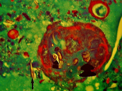
Fig.24 .- The arrows indicate the presence of typical corona hooks - which is located at the head of scolices-floating freely within the cyst, from scolices dead. Prof. Garfia.A


Fig. 25 .- The figure seems to correspond to the necrotic ghost of a daughter vesicle containing several scolices, which come from the numerous hooks contained in it (thick arrows). Outside of the gallbladder found floating in the magma hooks gel that fills the cyst. Note the appearance of hooks in cross section and the recess in its interior hook small arrowed. Prof.Garfia.A


Thanks .-
I am deeply grateful to Dr. Angel Prado Morales, Chief of General Surgery Hospital Ciudad de Coria, Coria City (Cáceres), for the invaluable assistance provided for the preparation of this case.


Bibliography
JI González Muñoz et al .- giant splenic hydatid cyst.
Cir Esp 2006; 79. nº 2. 


Thursday, March 24, 2011

Woman Showing Boobs Saree

Grease!


Jacket: Zara (very old)
T : Zara
Shorts: Vintage (Levi's 501 best-known )--->
blog me! jajajajaja.
Trousers: Calzedonia
Moccasins:
Zara Bag: Zara


The other day I went for beers with my friend Pilar (which appear in the photo .... that mássssss adore!) And there is no plan that I like better here Sevilla! As I started to get dressed and that stuff ... and now this ... I also think that this goes well and the end, without eating or drinking, I ended up with a full look at what " Grease "... or so it seemed to me! jajajja and the truth is that I liked bastanteos! =).
shorts, as I said above, are some old acquaintances blog in fact the previous post, but I love when I saw, I always come in handy for all the looks that come to mind ..... has happened to this once in a garment? is obsession! jajajajaj

also want to show the bag and shoes had my friend Pilar , because it was ideal as a whole, but the accessories are amazing! Both the bag and the shoes are her mother (mother-daughter typical robbery), a hoard that has for many years made a green skin more impossible ideal and original! and is new! That alegríaa, let that meeenamoré of them, indeed! =). You like it @ s?


cielines
Well that's it for today! Prontito we are here again!

PS: I tell you that this weekend I go to ... that ganassss nieveee! Landing and bring you photos of the mountain! jajajaja

Thanks for the comentariosss, I wish you a great weekend!

muaa


Marya *

Tuesday, March 22, 2011

How To Use Hp Built In Cam For Oovoo

96.3.-ECHINOCOCCOSIS IN FORENSIC PATHOLOGY IN SPAIN. PROF.GARFIA.A

96.3.-HYDATID CYSTS  IN SOUTHWEST OF SPAIN.  A FORENSIC POINT OF VIEW.  HISTOPATHOLOGICAL FINDINGS IN LUNGS AND LIVER.
Prof.Garfia.A

96.3-HIDATIDOSIS Patologia FORENSICS EN EN EL SURO DE ESPAÑA: HALLAZGOS Pulmonary Y HEPÁTICOS.
Prof.Garfia.A

96.3-hydatidosis IN FORENSIC PATHOLOGY IN SOUTHWEST SPAIN: PATHOLOGICAL FINDINGS IN LIVER AND LUNG.
Prof.Garfia.A

CASE NO 3
Case description
is an individual, a German national, who died in a tourist city on the Andalusian coast. The autopsy report gave a diagnosis of death from acute cardiorespiratory failure. Blood alcohol content was close to 3.0 g / L.

Histopathology
The liver showed numerous cysts, the largest of which measured about 5 mm in diameter, some of them were calcified and necrotic centers.
Microscopically, cysts were formed by a thick capsule of dense fibrous tissue, surrounding a cavity containing calcified debris and covered by a fabric composed of several cell layers within which were epithelioid-like cells and multinucleated giant cells (arrows). The cysts were found in different evolutionary stages towards the formation dense fibrotic nodules, and solid cystic cavity. None of the cysts examined was possible to display the membrane prolígera / germination, or the laminate layer, typical of hydatid cysts. It issued a diagnosis of liver cysts compatible with inactive hydatid cysts in the evolutionary process towards residual fibrosis. The remaining parenchyma showed steatosis, macrovesicular type, severe panlobular (+ 75% of hepatocytes affected), alcohol compatible.


Fig.1 .- Masson.Panorámica Hígado.Tricrómico of liver at low magnification. Inactive hydatid cysts. The composite photograph shows five cysts (three in the field below, one at 9 in the dial face, and another at 14 hours), which are at different evolutionary stages. Note that the cavity field below have a relatively large necrotic material occupied by calcium. The cyst located at 9 in the dial face, smaller size, has a coating endoquístico by a newly formed tissue containing epithelioid cells and multinucleated giant cells, foreign body replaces what would be the cystic lining or prolígera germination. Prof.Garfia.A


Fig.2 .- Detail inactive cyst, located at 9 in the dial face. Note the thick adventitial or pericyst, consisting of dense fibrous tissue oriented, the absence of laminated membrane, the membrane destruction germination or germ layer and its replacement by a multilayer cell containing fibroblasts, epithelioid cells and giant cells of Masson multinucleadas.Tricrómico. Prof. Garfia.A


Fig. 3.- Detail multilayer membrane replacement cell cystic germination, which has disappeared (inactive cyst). The arrow pointing to a giant cell multinucleada.Tricrómico Masson. Prof. Garfia.A


Fig. 4.- Detalle para mostrar la formación de células gigantes multinucleadas (flecha), en el seno la cavidad quística, encargadas de la fagocitosis de la membrana germinativa. Tricrómico de Masson.  Prof. Garfia.A


Fig.5. fibrotic cyst-Detail of Figure 1.
Prof.Garfia.A