93.-MUERTE IN CUSTODY immunocompromised patients: pulmonary pathology.
Prof.Garfia.A
Custody 93.-DEATH OF A POSITIVE IN AIDS DRUG ADDICT: PULMONARY FINDINGS. Prof.Garfia.A
Background
Among the subject's background was a history of drug addiction-a-long-standing abuse.
Two weeks before his death he had returned from a therapeutic community in which it was admitted for treatment for addiction.
Circumstances of death
The body was found in the supine position, lying on the bed cell in a police station, where he was detained. It is, therefore, the death of a detainee or called in forensic medicine, death or custody.
Such deaths have a special interest in legal medicine because it occurs in individuals who are detained in jail or at the police station and often lead to family complaints against the police, on suspicion of abuse, concealing the desire-often-to get more compensation than a clarification of the cause of death.
; Most of the time these deaths are a relief, rather than a setback, consciously or unconsciously, for the families.
; Most of the time these deaths are a relief, rather than a setback, consciously or unconsciously, for the families.
family contributions
According to relatives, the subject "suffering from nerves" without specify more data. Of course, commented that he was diagnosed with pulmonary tuberculosis and bronchitis, so it was treated with "Ventolin".
We do not know if, in addition, was treated with antibiotics.
We do not know if, in addition, was treated with antibiotics.
autopsy examination
At autopsy there was little consideration of relevant medical data, except pulmonary findings. Described bilateral pulmonary adhesions and the presence of abundant calcifications of variable size, which affected all lobes of both lungs.
HISTOPATHOLOGY
Macroscopically, cuts had multiple lung nodules chalky, white, with a necrotic center caseum and looking, of varying size.
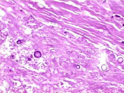
Microscopically were well defined and were defined by a fibrous capsule. In the center of the nodules, in addition to necrosis, there were areas of calcification and, more peripherally, spherules of different sizes.
Fig.1 .- Macroscopic lung, cut.
Note the presence of numerous "nodes" of varying size that are surrounded by a fibrous capsule, which has a slight bluish color in the picture. The section offered a look nodules with necrotic lesions, caseous, white and chalky. On the periphery of the lesions could be found few multinucleated giant cells and minimal inflammatory response, cell linfoplasmocitarias.En the rest of the lung parenchyma parenchymal foci were detected in nodular lymphoid tissue. staining for bacilli Koch was negative and the periphery of nodules near the capsule, we found spherules of variable size (5 to 30.40 microns or so.).
Prof. Garfia.A
Since the case was presented at the "Forum of Pathology Club autopsy Autopsy" I belong ,
(http://eusalud.uninet.edu/cl_autopsias/Casos/10.03/caso.htm) will incorporate the comments made by members of the Club.
Comments on Case .-
1.- Dr. E. Moro said:
seems a form of pulmonary cryptococcosis. A case of Crytococcus neoformans meningitis appeared also here some years ago:
(http://eusalud.uninet.edu/casos/caso42.html).
2 .- Dr. E. Mayayo wrote:
very interesting case and crumb. I do not think a cryptococcosis as noted by Dr. Moro. I lack the characteristic mucoprotéicos halos around the spores, which give an appearance of sun. Also, the fact that they like to have a diversos.Me sizes and staining Groccot with mucicarmine. I am inclined to spherules of calcium may be because there are very dense areas in the center of the nodules. Von Kossa staining can be very useful and enlightening. The silicotic lymph nodes remember me but the question is what do in a patient of this kind? I do not think it is due to material accompanying the adulteration of drugs because they are too big. And we will open up the door dianóstico can be very enlightening as all cases are being presented.
Dr. 3.-comment "Rokitansky"
Macroscopically, it may well think of a tuberculosis. The "caseous necrosis", however, also seen in other circumstances, for example, microscopic images show infections micóticas.Las fibrocaseosas nodular lesions with calcification and no apparent inflammatory response, probably, the fact is due to the time evolution so long of nodules (lesions "plus chronic), and the subject's immune status (immunocompromised).
The spherules located near the capsule fibrous "are perhaps the expression, if somewhat distorted, of a fungal infection early, or simply try to deposits of calcium salts in the context of dystrophic calcification?
Luis Thompson and David Oddo make a review of the "Fungal Opportunistic Mycoses" on the following page:
(http://edcenter.med.cornell.edu/CUMC_PathNotes/Respiratory/Respiratory.html).
Figs. 2.3 .-
Cortes lung stained with PAS. Discrete nodules show positivity. Prof. Garfia.A
Figs. 4, 5 and 6 .-
correspond to progressive increases in the interior of the nodules to highlight the PAS + spherules that are located near the capsule. The peripheral rim of the spherules is PAS positive and the picture No. 5 is located at nine of the dial face, a small spherule body size has a central dense, PAS +. staining (silver staining) of Von Kossa for calcium was positive demonstration. Prof.Garfia.A
Fig. 7. -
Photography, a small increase in lung stained with Grocott technique for demonstration fungus shows negativity for the presence of those. Prof.Garfia.A
Fig. 8 .- Histological section stained with Masson trichrome. In the center of some nodules appeared necrotic material dyed red. With the technique clearly demonstrates the nodular fibrous capsule. Prof.Garfia.A
Diagnosis, comments Authors, cause of death and bibliography .-
1.-Diagnosis .-
fibrocaseosas Injury, old, negative BK, with calcifications dystrophic, suggestive of tuberculosis in immunocompromised patients.
Comments .- 2.-
The comments from some participants in the Forum regarding the diagnosis of case I seem very successful.
Cryptococcosis Diagnosis requires the presence of characteristic mucoproteicos halos around the spores, as indicated by Dr. Mayayo. Stains for fungi were negative. Moreover, the size of the spherules as disparate, their proximity to the fibrous capsule and the presence of other fully calcified nodules, induced a suspicion that the spherules were treated dystrophic calcium act as initial crystallization centers.
3 .- cause of death .-
Chemical analysis showed the presence in blood of toxic following:
3.1.-Cocaine and its metabolites.
3.2.-toxic levels of methadone.
3.3.-important concentrations of anxiolytic.
Death was attributed to acute poisoning by drugs of abuse (polydrug use), where methadone played a crucial role taking into account also the possibility of synergies and enhancing the effects of call, in Spain, "rebujito narcotic."
Disclaimer author .-
The rebujito is the popular term applied to the common drink consumed at the Fair Sevilla. It consists of a mix of "Seven Up and Manzanilla, which makes the most refreshing chamomile and tolerable, and at that time usually does Fair enough heat.
BIBLIOGRAPHY
1.-View Dr. Rokitansky comments on the locations of interest, on the Web, for lung disease.
addition, it interesting to the knowledge of two other appointments:
2.-Karch SB .- The Pathology of Drug Abuse. NYCRC.1996.
3. - Drummer OH .- The Forensic Pharmacology of Drugs of Abuse. Arnold Publishers.London.2001.Especialmente interesting to all who may be interested in the interpretation of analytical results and postmortem drug concentrations in relation to the cause of death.


0 comments:
Post a Comment